The classical sonographic method to image the abdominal aorta (AA) is the anterior mid-abdominal transverse and longitudinal approach (Fig 1 and 2) using the curvilinear, low frequency probe.
However, there is another method of looking at the AA; the right lateral intercostal approach.


Fig 1 Mid abdominal transverse approach


Fig 2 Mid abdominal longitudinal approach
How often, especially in the obese or tender abdomen, have you found it difficult to visualise the aorta with ultrasound?
We try everything we can: asking the patient to bend their knees to relax the rectus muscles, applying gentle graded pressure to the probe in an attempt to displace the gas which is often in the way. Most of the time we barely visualise the AA, and can’t say much other than excluding a big AAA.
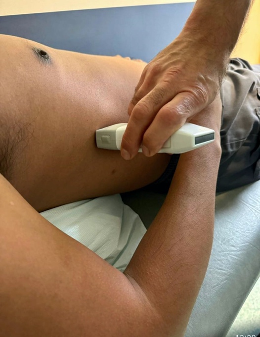
If we want to be more accurate, we can try the right intercostal approach (Fig 3); utilized to study the inferior margin and hilum of the liver, the gallbladder and the IVC, tilting the probe towards the IVC. This is particularly useful with imaging the first 10-12 cm of the AA.


Fig 3 Right intercostal approach
Some of the aforementioned obstacles (air in particular), are removed with this view. In fact, the liver and the IVC (that is located above the AA on the screen because anatomically it lays to the right of the AA) act as main acoustic windows and no pressure is exerted on the abdominal viscus because the probe is applied over the rib cage.
The difference between this view and the classical anterior view is that we see the AA longitudinally and “under” the IVC instead of at the right side of it.
Clinical case
A 44 year old man, who presented to the ED for central chest pain and with negative ECG and troponin, was referred to the Same Day Emergency Care Unit (SDEC) the following day to rule out pulmonary embolism.
Tachycardia and epigastric tenderness were the only findings at the time of presentation in the SDEC, so, besides a focussed echo study, we decided to assess the AA with POCUS .
The traditional anterior approach allowed us to exclude an AAA and provided images that raised the suspicion of an intimal flap within the AA (Clip 1).
This was confirmed with a clearer view of the AA (and of the intimal flap) obtained with the right longitudinal intercostal approach (Clip 2).
A flap was confirmed within the descending thoracic aorta using the suprasternal approach with a phased array probe (Clip 3).
Diagnosis
A Type B aortic dissection was confirmed on the CT aortogram.
Conclusions
An alternative US perspective such as that given by a right intercostal approach can improve the visualisation of the abdominal aorta, making it easier to diagnose an intimal flap in the context of aortic dissection.