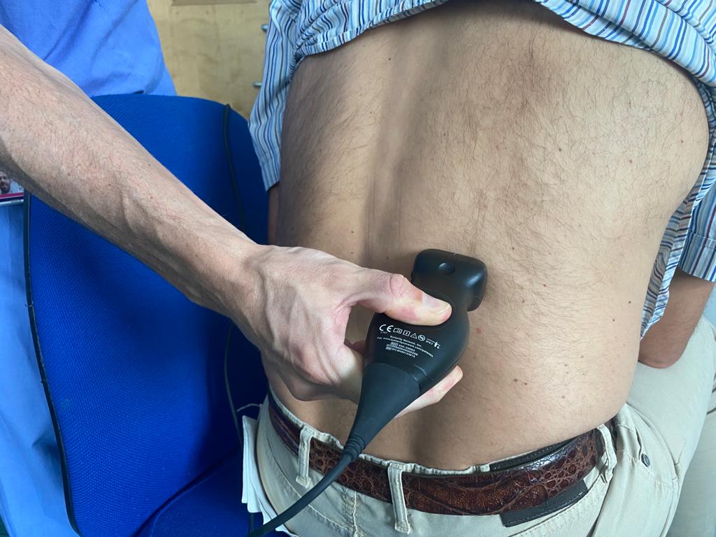
Gently hitting the costophrenic angle to elicit kidney pain is common practice in patients with suspected renal colic or pyelonephritis. Have you ever thought of doing it with an ultrasound probe?


Clinical case
A 29 year old lady presenting with fever spikes associated with back pain and severe headache was admitted in the Acute Admission Unit. She localised the pain at the level of the right costophrenic angle.
Differential diagnosis
Pyelonephritis was initially suspected and treated but the reported pain could not be elicited by palpating the area where the right kidney was meant to be (negative Giordano’s sign).
To check whether the pain was really originating from the kidney I then decide to use an ultrasound probe. Once identified the right kidney using a posterior approach and with the patient in the sitting position, I gently patted with the probe exactly over the organ (let’s call it sonographic Giordano’s manoeuvre) but the patient didn’t report that area to be the most tender.
Using then a lung pre-set and sliding further up to look for possible causes in the lung it appeared clear that the source of all the problems was there. It was indeed when I gently patted the area above the consolidation identified in the right lower lobe (see the “shred sign”, air bronchogram” and the B lines) that the patient reported the sharpest pain.
Video: Sonographic Giordano’s Sign:
Diagnosis
Pneumonia
Final comments
Point of Care Ultrasound (POCUS), by identifying the exact location of each organ and allowing for focused pressure, can be useful to identify the source of symptoms during physical examination.
We have seen it applied with pain of suspected origin from the kidney.
Another very common application is in suspected biliary colic or cholecystitis, the so-called sonographic Murphy sign (see video).
Video: sonographic Murphy’s sign:
POCUS allowed to stop the ACS treatment and refer to the surgeons this patient with cardiac stents presenting with central chest pain, however different from the angina he suffers from, negative troponins and positive sonographic Murphy sign !
A non-dilated gallbladder, although replenished with stones, without signs of cholecystitis would have not received too much attention in this context if it was not still painful under the probe (after a one hour long biliary colic).
Very rarely I have seen this last sign reported on a “formal” ultrasound study (never the Giordano). This is why we should all learn POCUS!
